Surgical Repair of Atrioventricular Septal Defect Dimas Yusuf
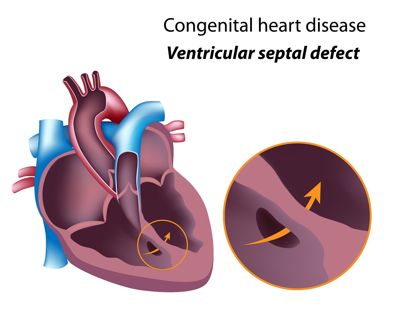
Ventricular Septal Defect (VSD) A ventricular septal defect (VSD) is an opening in the interventricular septum, causing a shunt between ventricles. Large defects result in a significant left-to-right shunt and cause dyspnea with feeding and poor growth during infancy. A loud, harsh, holosystolic murmur at the lower left sternal border is common.

Pin on Cardio
A ventricular septal defect (VSD), is an abnormal opening between the right and left ventricles. It can vary in size, and when they're small they can sometimes close on their own in that first year of life. It's when they're on the larger side that they start causing significant problems for our patients.

Atrial Septal Defect, Normal Heart, Infancy, Pulmonary, Arteries, Atrium, Asd, Early Childhood
A ventricular septal defect (VSD) is a congenital heart defect. This means that your baby is born with it. A VSD is a hole in the wall (septum) that separates the 2 lower chambers of the heart (right and left ventricles). VSDs are the most common type of congenital heart defect. The heart has 4 chambers: 2 upper (atria) and 2 lower (ventricles.

an image of the human heart with labels
Outline Lesson Objective for Congenital Heart Defects Understanding Congenital Heart Defects: Gain comprehensive knowledge about the various types of congenital heart defects, their anatomical and physiological implications, and the impact on cardiac function. Identification of Common Defects:

Ventricular Septal Defect (VSD) Atrial Septal Defect (ASD) childrenhealth children health
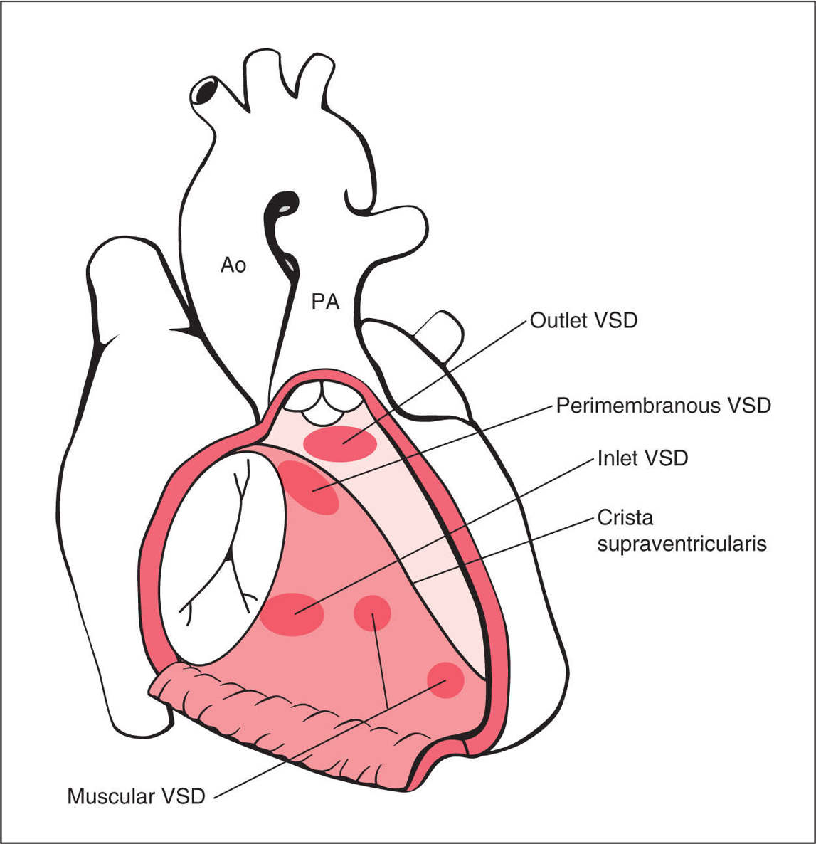
Ventricular septal defect (VSD) is the most common congenital cardiac anomaly in children and is the second most common congenital abnormality in adults, second only to a bicuspid aortic valve. An abnormal communication between the right and left ventricles and shunt formation is the main mechanism of hemodynamic compromise in VSD.
Congenital heart disease ventricular septal defect The Pulse
The atrioventricular septal defect is a congenital cardiac malformation that is characterized by a variable degree of the atrial and ventricular septal defect along with a common or partially separate atrioventricular orifice. [1]
Ventricular Septal Defect The Patient Guide to Heart, Lung, and Esophageal Surgery
What is a ventricular septal defect (VSD)? It's a congenital heart defect that occurs when there is a "hole" in the ventricular septum. This causes an increase in blood flow to the lungs. Quick Facts about VSD 1 in every 240 babies born in the United States each year are born with a ventricular septal defect.

Ventricular Septal defect Symptoms, Causes & Risk Factors Dr Raghu
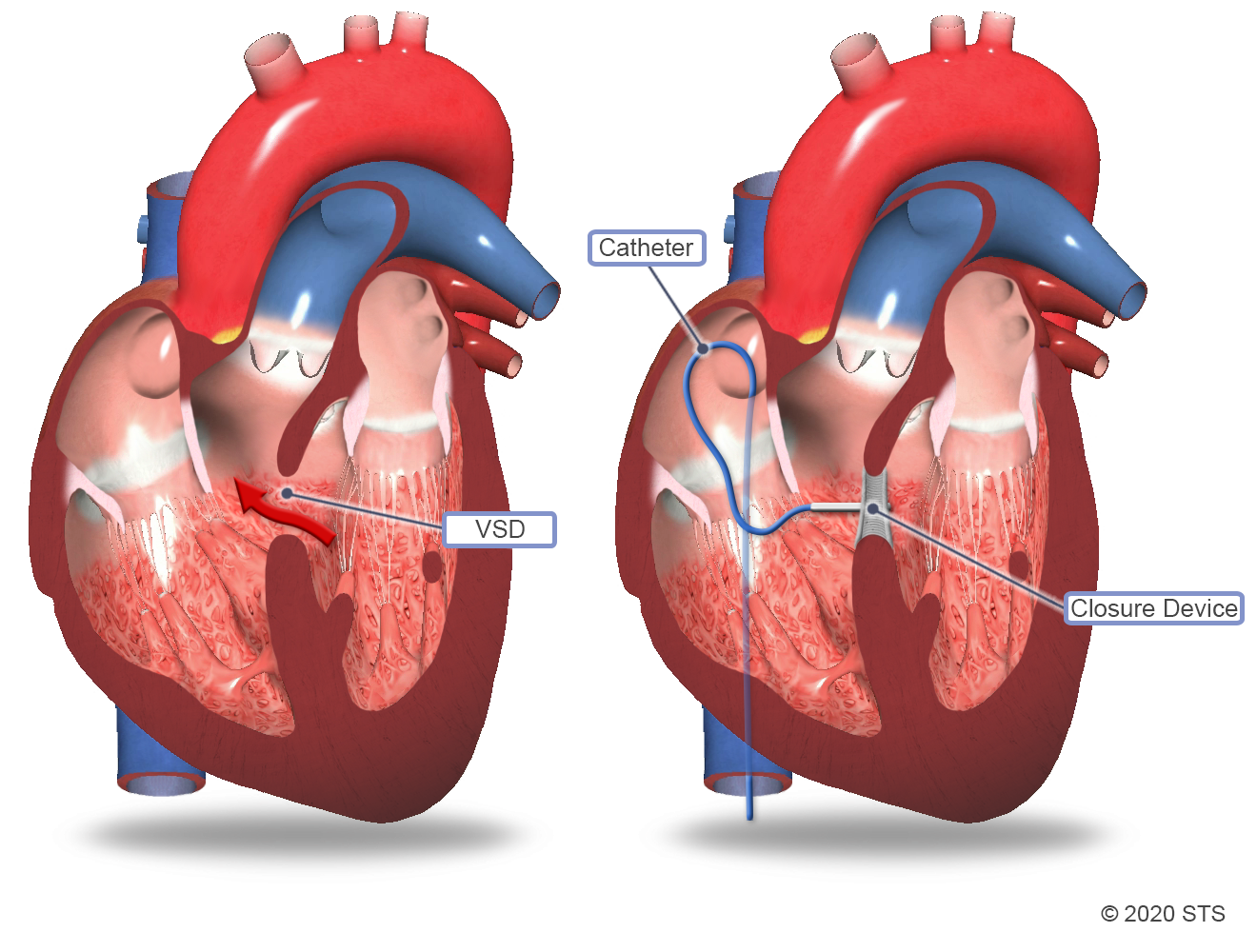
The present article describes the clinical aspects of ventricular septal defects and current management strategies. Ventricular septal defect (VSD) is a common congenital heart defect in both children and adults. Management of this lesion has changed dramatically in the last 50 years. Catheter-based therapy for VSD closure, now in the clinical.
/GettyImages-141483002-5bb39d7746e0fb0026439ac9.jpg)
What Are Ventricular Septal Defects?
Ventricular septal defect (VSD) nursing NCLEX review over the pathophysiology, signs and symptoms, complications, nursing interventions, and treatments.What.
What is Congenital Heart Disease Types, Causes, and Symptoms
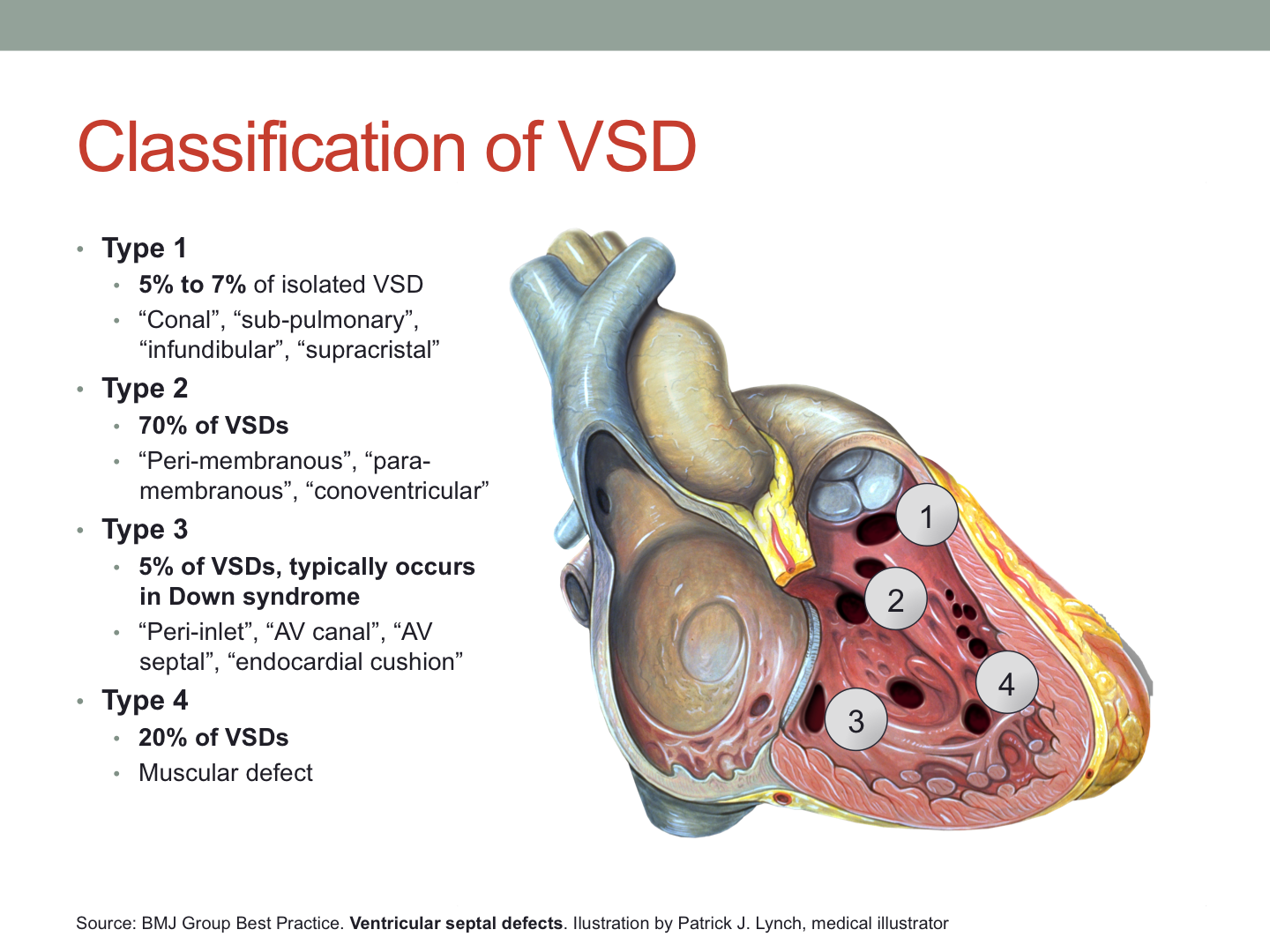
The indications for closure are moderate to large VSDs with enlarged left atrium and left ventricle or elevated pulmonary artery pressure (or both) and a pulmonary-to-systemic flow ratio greater than 2:1. Surgical closure is recommended for large perimembranous VSDs, supracristal VSDs, and VSDs with aortic valve prolapse.

Pin on Nursing stuff
Ventricular Septal Defect is a congenital heart anomaly characterized by a hole in the wall (or septum) separating the lower chambers of the heart or the ventricles. This causes oxygen-rich blood to travel back to the lungs rather than be pumped out to the entire body. Notes on VSD:
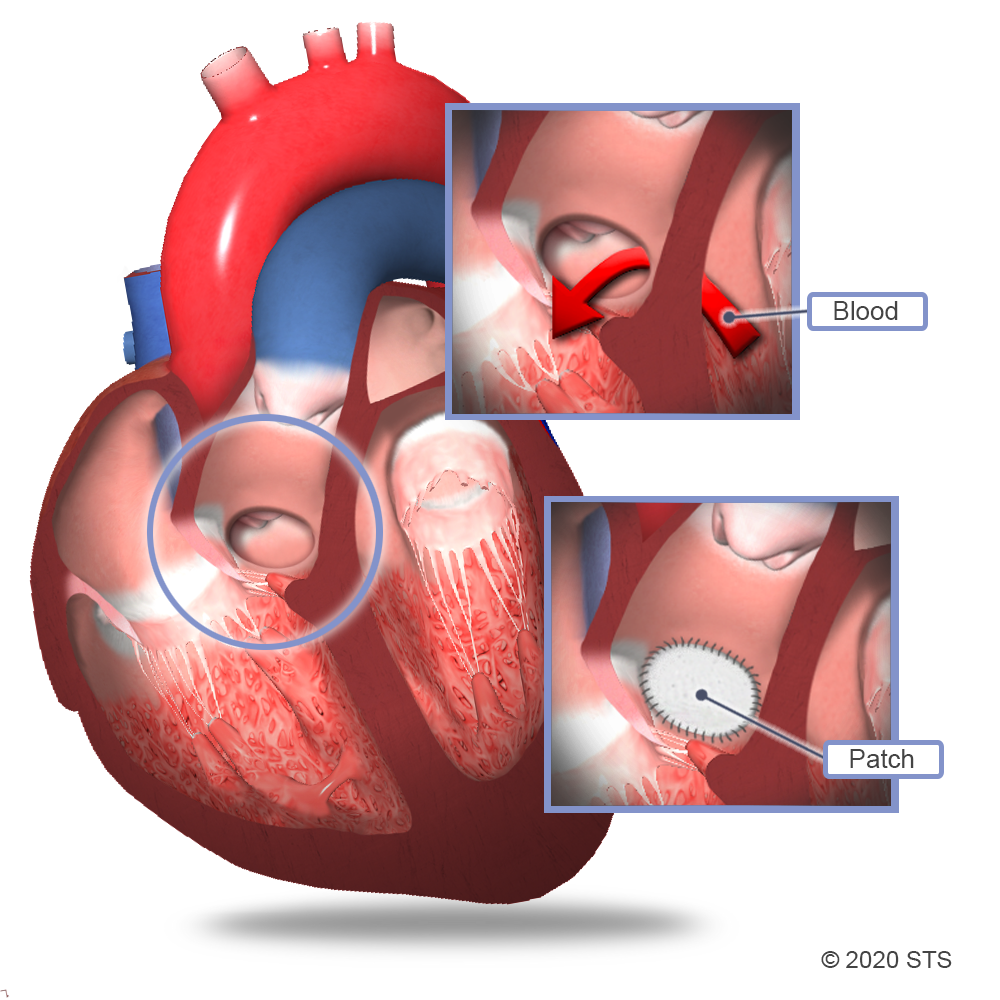
Ventricular Septal Defect Surgery The Patient Guide to Heart, Lung, and Esophageal Surgery
Ventricular septal defects are defects in the interventricular septum that allows shunting of blood between the left and right ventricles. Usually congenital, but rarely acquired after myocardial infarction or trauma. May be associated with other congenital defects such as tetralogy of Fallot. Significant left-to-right shunting results in.
Atrial, Ventricular, and Atrioventricular Septal Defects Obgyn Key
1. Managing Decrease in Cardiac Output 2. Promoting Effective Family Coping 3. Improving Tolerance to Activity 4. Preventing Injury and Infection 5. Administering Medications and Providing Pharmacological Interventions 6. Providing Perioperative Nursing Care Recommended Resources See also What is congenital heart disease?
Ventricular Septal Defect Surgery The Patient Guide to Heart, Lung, and Esophageal Surgery
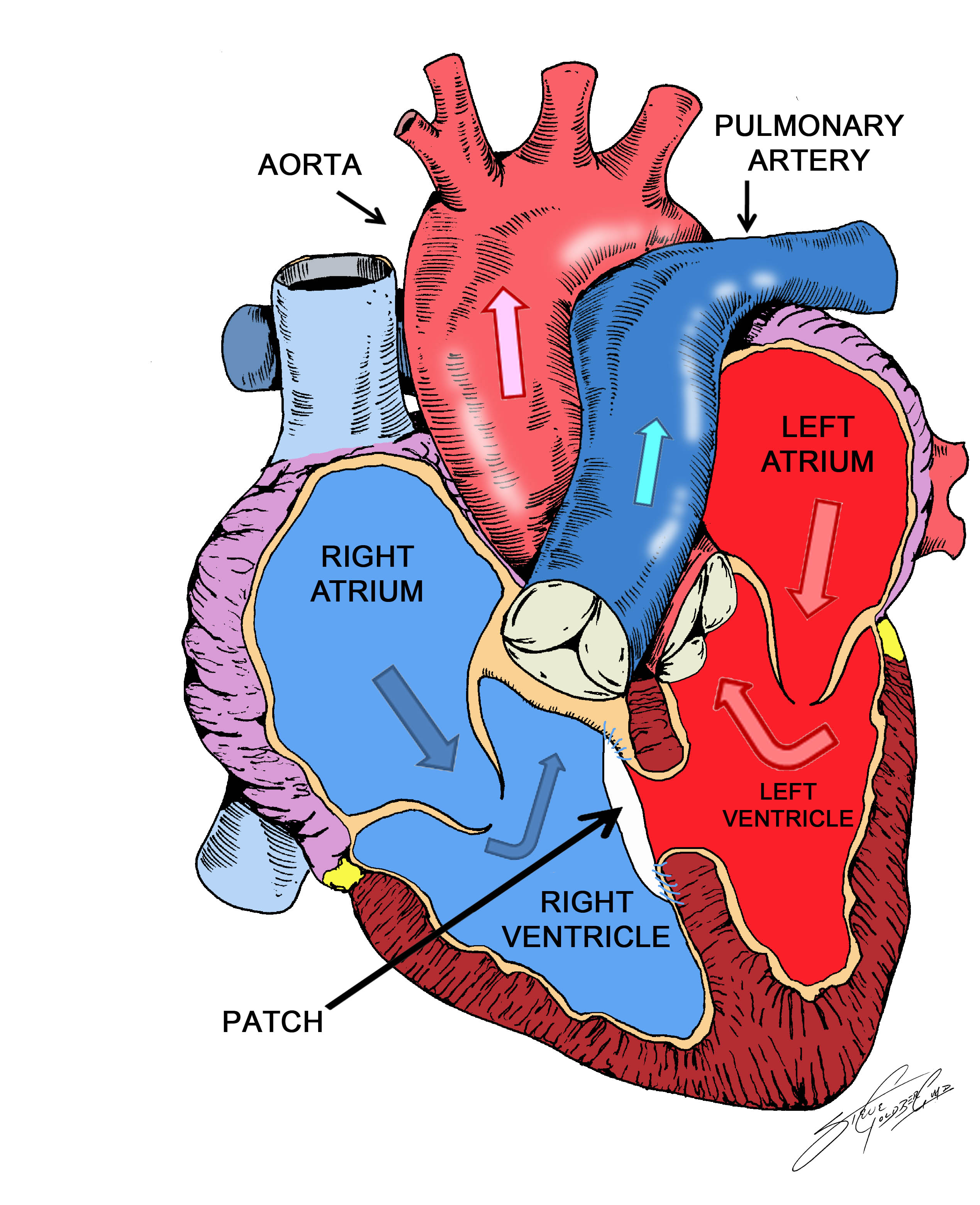
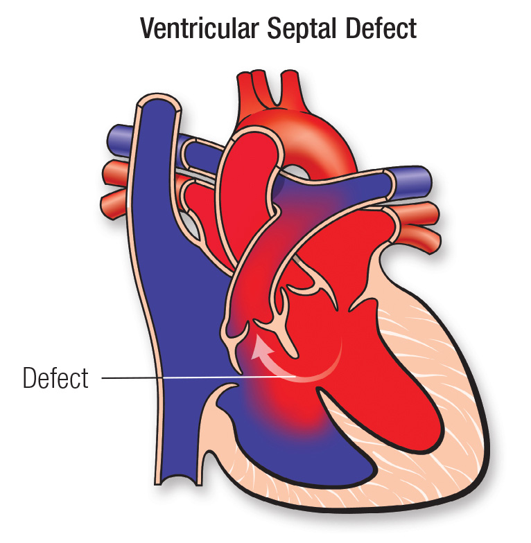
A ventricular septal defect (VSD) is a hole between the right and left pumping chambers of the heart. The heart has four chambers: a right and left upper chamber called an atrium and a right and left lower chamber called a ventricle. In the normal heart, the right and left chambers are separated from each other by a wall of muscle called a septum.
Ventricular Septal Defect (VSD) Seattle Children's
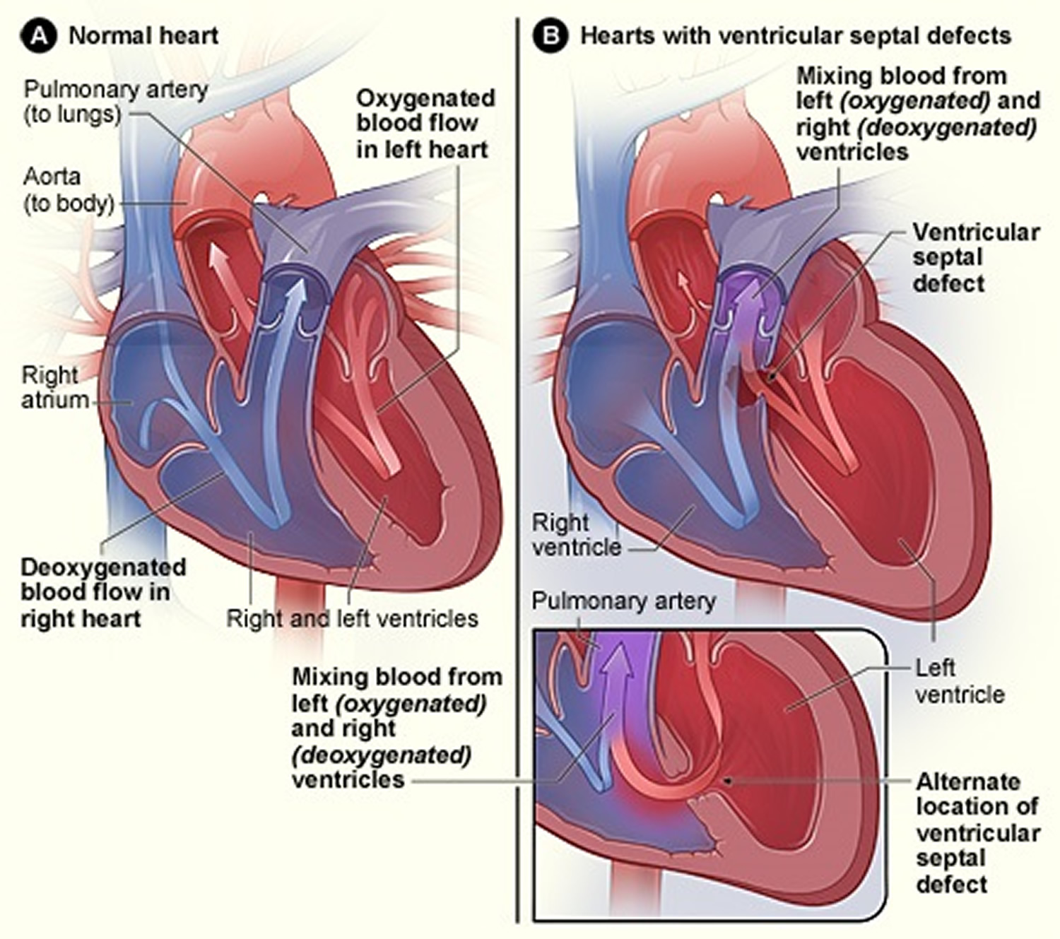
A ventricular septal defect changes the direction of blood flow in the heart and lungs. The hole lets oxygen-rich blood go back into the lungs, instead of going out to the body. Oxygen-rich blood and oxygen-poor blood now mix together. If the ventricular septal defect is large, the blood pressure in the lung arteries may increase.

Heart Defects Ventricular septal defect, Atrial septal defect, Cleft lip and palate
Treatment Self care Coping and support Preparing for your appointment Diagnosis Some ventricular septal defects (VSDs) are diagnosed soon after a child is born. However, ventricular septal defects (VSDs) may not be diagnosed until later in life.